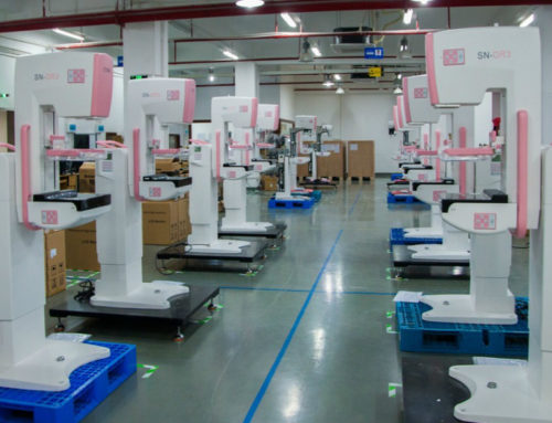
Breast cancer is the most commonly diagnosed cancer in women – second only to skin cancer. Nowadays the mainstream detection methods include full field mammography, mammography ultrasound and mammography MRI,and the new technology named cone beam breast CT which may be the possible detection in the near future.
Now the full field mammography is the most popular detection method for the most women,replacing the outdated film analog machine in the past decades.And recently the newly launched 3D mammography ,also named digital tomosynthesis mammography is developing rapidly,becoming the latest innovation in breast cancer screening technology, and it is already showing promising results
Many international brands are developing its own model of 3D mammography , such as Hologic , Planmed and Fuji Film,etc,and have been occupying certain high-end markets. You can check the list of manufacturer of 3D mammography : updated 2017
What is 3D mammography and how does it work?
Breast tomosynthesis, also called three-dimensional (3-D) mammography and digital breast tomosynthesis (DBT), is an advanced form of breast imaging where multiple images of the breast from different angles are captured and reconstructed (“synthesized”) into a three-dimensional image set. In this way, 3-D breast imaging is similar to computed tomography (CT) imaging in which a series of thin “slices” which here is 1mm thickness of enhanced image are assembled together to create a 3-D reconstruction of the body.
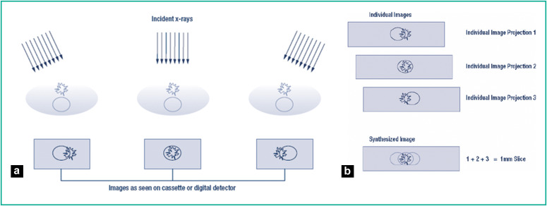
Very much like traditional 2D mammograms, 3D mammograms work by taking x-ray pictures of breast tissue, which are then analyzed by a radiologist.
But, unlike conventional X-ray imaging, 3D mammograms have an additional x-ray robotic arm that rotates around the base of the machine, capturing multiple images in a very short amount of time.
Let’s take Hologic® – Selenia Dimensions™ mammography systems for example.As a leading supplier of 3D mammography, Hologic launched the 3D tomosythesis mammography 3 year ago The X-ray tube of this model continuously sweeps in a 15-degree arc over the breast to acquire a series of low-dose projection images at multiple angles in less than 3.7 seconds. The whole process take 15 photos with each 1 angle for 1 picture.
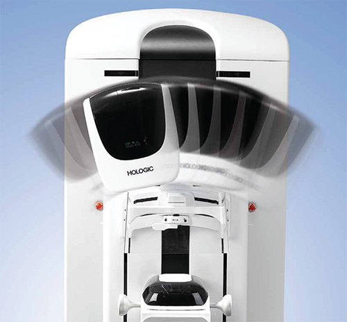
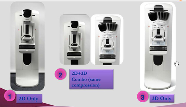
Hologic Selenia mode
And currently the machine could support 3 modes as above which are 2D only , 2D+3D combo and 3D only.With 2D mode,the machine is used just as standard digital mammography with common four position taken and,with the combo of the 2D and 3D, the C-arm would take make a pause on the common vertical and 45 °position,taking shoots of breast besides getting 3D images.Then the images are then digitally projected onto a computer, be combined and reconstructed into a 3D image,which fianlly would be analyzed both by the computer’s algorithm and the radiologist.
What is the the advantages of 3D mammography?
Conventional 2D mammograms use x-rays to capture a limited image of breast tissue of certain position while 3D tomosythesis mammography takes multiple and continuous images ,which can be observed one by one or layered to produce an entirely three-dimensional view of the breast tissue. Compared with the 2D mammography ,3D mammography features:
1.Higher resolution benefits the detection accuracy
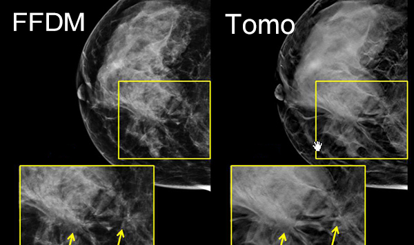
Generally speaking, 3d mammogram are able to show at most possibility the overlapping part of breast parenchyma and greater accuracy in pinpointing the size, shape and location of breast abnormalities,which leads Less chances of false positive and avoid vagueness of small parts at most degree.
According to the study of Friedewald SM, Rafferty EA, Rose SL(credit)3D mammography has been shown to improve breast cancer detection by 27-50%.Combined 2D/3D (digital breast tomosynthesis) mammography is superior to 2D mammography alone in the diagnosis of breast cancer, as shown by a number of large studies. One large U.S. study of 454,850 patients found that combined 2D/3D screening detected 5.5 cancers per 1,000 women, compared with 4.3 cancers for 2D screening alone—a 28% higher detection rate and a 16% lower recall rate.
Credit:Friedewald SM, Rafferty EA, Rose SL, et al: Breast cancer screening using tomosynthesis in combination with digital mammography. JAMA 311:2499-2507, 2014.
According the American College of Radiology;the image performance of breast cancer are frequently indicated by main factors following: tumor, calcification, partial mammary glands,breast burr .With DBT technology, not only the size,shape and border, quantity of the lump,also the status of calcification situation around the tumor could be viewed much clearly than FFDM. Detailed speaking ,it helped
- Increase the detection accuracy in the patientswith GLAND BREAST ISSUE
With 2D mammography, the wrong detection of patients with gland tissue may reach high up to 76% as the tissue is so dense to cover the tumor. While through tomosythesis, it would be easier to notice the location and shape of tumor with a 3D image ,which does increase the accuracy in early detection phase.
- Higheraccuracy in showing the quantity and border of lump
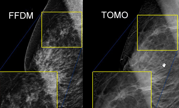
3D images could not only help to find the small tumor ,but also could depict the border of tumor,which could not be detected in FFDM machine because of the overlapping tissue existence.The successful finding of the small size tumor and the existence of burr could have increase the finding rate of the magliance of the tumor,which as a result ,could help a lot in finding a more effective curing method in the future.
- Better diagnosis of false positive
In the shooting process in in FFDM or analog film screen mammography,X ray is hard to go through some dense breast issue area so it may be seen as suspicious focus,especially the in the gland tissue.They are sometimes proved to be a false positive in the following development of the tumor,which would do great harm to the treatment .The DBT do a great function in eliminating the possibility of this kind of false positive diagnosis.
Using this method increases the number of early defections and significantly lowers the number of patients that have to retake mammograms due to unclear or suspicious results. Fewer call backs. 3D mammography has been shown to lower recall rates by 17-40% according to the reports.
2.Reasonable dose radiation
Compared with 2D mammography, Digital tomosynthesis screening takes a few seconds longer. Nevertheless, the slightly increased time of screening does not imply that the amount of radiation to the patient also increases. In fact, 3D mammograms have a lower radiation dose than traditional analog mammography. Please kindly check the comparison chart below:
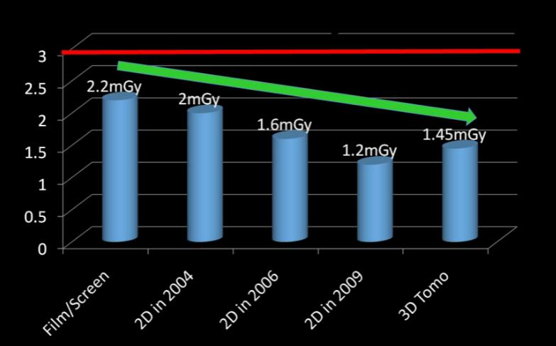
Though the whole amount of dose use of 2D + 3D is more than 2D machine only, It is complied with MQSA standard by FDA which is average rate is no more than 3mGy.
And to decrease the dose radiation as much possible, some supplier like Hologic use the so-called synthetic 2D reading. For this reason, Hologic developed C-View™ software, which generates the 2D images directly from the 3D dataset and avoids the need for the 2D exposures altogether.
3.Relatively fast and comfortable detection process
The whole process takes no more than 10-15 seconds and just like the current mammography, the patient could stay one position releasing the trouble of moving frequently
A 3D mammogram screening applies a significantly lower amount of pressure on the breast than conventional 2D X-ray screening, which increases the patient’s comfort during the exam, and the precision with which the tomosynthesis units capture and reproduce the image.
Current development of 3D tomosynthesis
Global Mammography Innovations
Nowadays,3D screening technology has been under fast development worldwidely,starting from the academic study to clinical evaluation gradually.
Major medical device and technology manufacturers such as Philips and General Electric are focus on these high-end market,implementing 3D technology into their mammography machines and have been improving the functions continuously.
General Electric’s flagship model,Senographe Pristina™ mammogram now has a 3D variant with improved image quality and a new, ergonomic design, making the screening easier for patients, technicians, and radiologists.
MicroDose SI is the latest mammogram from Philips. Their newest tomosynthesis machine has an increased operating speed and dose efficiency without sacrificing image quality. Collaborating with radiologists, technicians and patients resulted in a design that caters to the patient’s comfort.
Apart from these two major medical device manufacturers,more companies are investing in mammography research and producing new and improved tomosynthesis machines. Some of the more notable companies and their respective tomosynthesis units are:
- Hologic®- Selenia Dimensions™ mammography systems
- FUJIFILM – Amulet Innovality
- Neusoft® – NeuCare Mammo DR™
- Planmed – Clarity 3D
- AMICO X-ray Technologies – Mammo-RPd
While regretfully,the 3D tomosynthesis machine is still applied normally on the academic field
as it would take relatively more time for doctors to learn and analysis,and besides,the high price scare off makes it hard to be used widely in common diagnosis cases.
3D Mammography at SinoMDT
As leading manufacuter of digital mammography,SinoMDT always been dedicated on the development of
new generation mamography,including the 3D tomosynthesis machine.With the support of local government,The new 3D generation XP-300 is under hot development and hopefully it would come out within 2 years.
It would might feature:
·Significantly reduced vibrations of the C-arm isorotation center
·Stable movement and easy installation process
·Smart, multi-level flexible compression
·Support for A-se and A-si detector panels
·Up to 15 images supported by the tomosynthesis function
·Additional post-processing equipment
If you would like to know more ideas and development of 3D mammography of Sinomdt,Please kindly contact with us.


